This is a long post. Today was not a normal day. It started as a normal day, we got up and went out for a walk. We went to the park and we went to the fields and we went to school. But then stuff became suspicious when I was put on the table to be the center of attention. I know I am interesting, but I am not sure the color of my gums is, or the size of my lymphnodes...
 |
| Clinical examination |
Then I was cannulated. They came to me with a machine that is scary and goes BZZZZZZ and takes fur off. That sound was the hard part. The easy part is when they put a tube into my arteries and blood came out.
 |
| They put a bandage to hide the cannula, maybe they were ashamed? |
 |
| Well, they should be! |
First they put some antibiotics in the cannula (ampicillin).. Then they put two more liquids, medetomidine and butorphanol. These are very boring liquids. So boring, that I fell asleep almost immediately. I barely had the time to hear Jen say "good night" as I let my head go to the floor. To be sure I stayed asleep, a little bit of propofol was given, but not too much, because I wasn't breathing very much and the other liquids worked very well on me.
I was intubated, shaved more and brought to the surgery room.
There I was put in dorsal recumbency and attached (just in case I decided to leave in the middle of the surgery?). My tube was hooked up to a gas-breathing system with inhalant gas (isoflurane) and to a series of monitors (ECG, capnograph...).
 |
| Dorsal recumbency |
 |
| Cleaning my belly |
 |
Left: Me
Middle back: TV for laparoscope
Middle front: Inhalation anesthetic device
Right: Monitors |
|
|
The red, yellow and green are the ECG probes.
The tube in my mouth is to keep me asleep. |
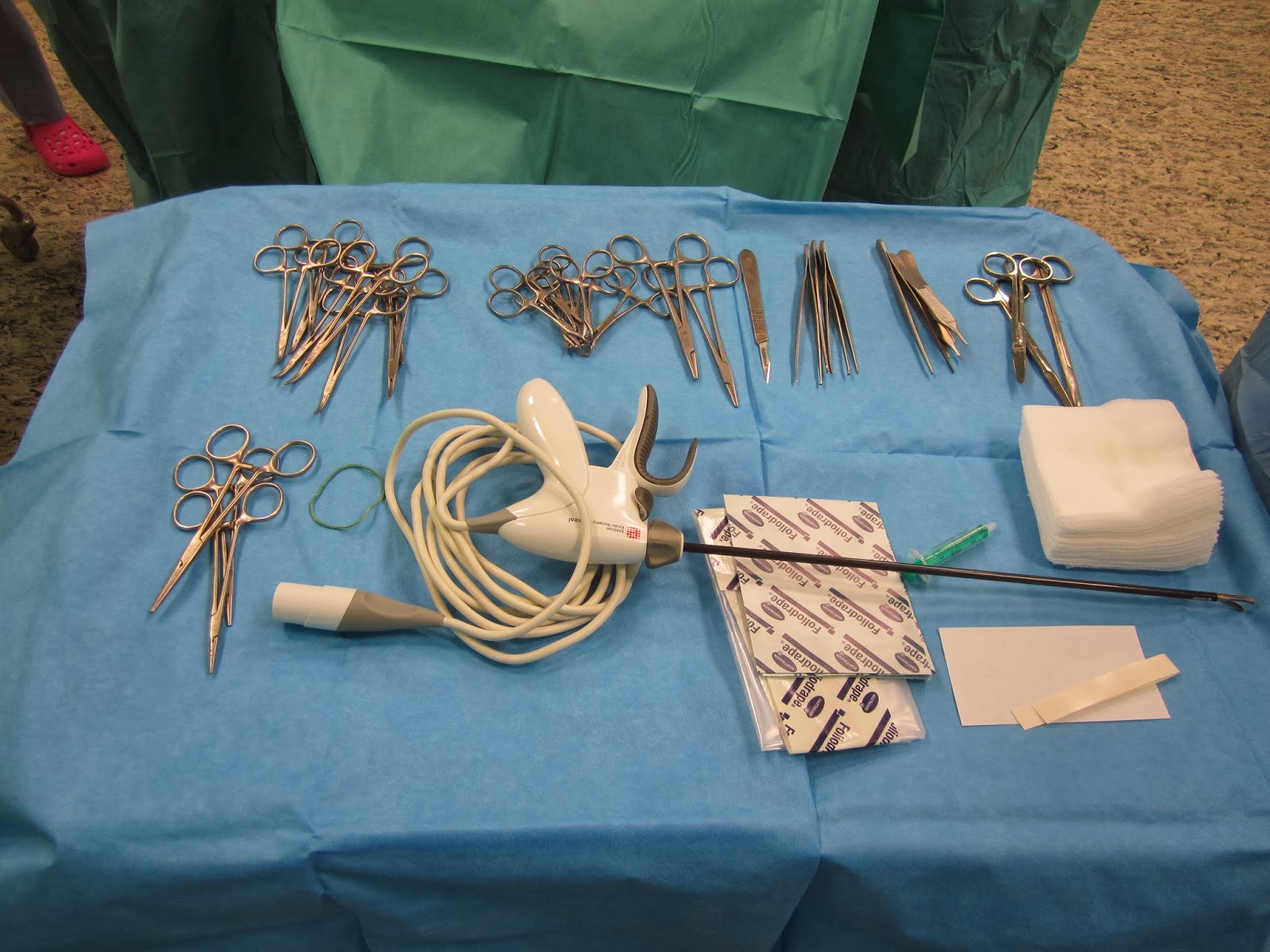
Then Dr. Crha prepared the instruments. It's not as easy as it looks, you must prepare for all possibilities that will occur during the surgery. Also, everything must be sterile. But Dr. Crha is a very good doctor, he knew what he was doing.
The goal of the surgery was a
spay (ovariectomy = removal of the ovaries) to prevent pregnancy and reproductive tract diseases, and a
gastropexy (suturing the stomach to the wall to prevent a condition called
Gastro-dilatation-volvulus).
Procedure:
- make 2 holes to install camera and grabbing tool
- find and catch ovary
- fix the ovary into position
- cut off ovary
- repeat steps 2-4 with the other ovary
- find and catch stomach
- suture it to the wall
- check everything is fine and close up
Oariectomy
Then the surgery started. I don't remember anything. First they made a hole in my abdomen. There are a lot of layers. Outside there is the skin (and subcutis), under that there are several muscles (but they cut in the middle of my belly, so it was only tendon tissue) and under that there is peritoneum, which is the inside wall of the abdomen.
Except that the poor surgeon was not lucky, and probably went to the exact same place that my stick injury happened (
remember, it happened like this). So it was a little harder than usual to get to the peritoneum. Usually, you use a trocar and pierce through, and feel two "POC" sounds: the first is the subcutis and tendon sheets, and the second is the peritoneum.
If the tube is not properly in the peritoneum, there will not be space to insuflate CO2. There is a little test you can do: put drops at the edge of the hole, and move the belly around a bit. It should make a bit more space inside the abdomen and suck in the droplets. If not, it means you only went down one "POC" and you are still in the out layers (subcutis).
So here are the 3 steps again:
 |
| Very small incision in the skin |
 |
| Pierce through with trocar POC-POC |
 |
| Put in CO2 |
They got in to the right place! The abdomen got bigger and bigger with air - I mean CO2!
 |
| The hand is taping on the abdomen and it makes an empty sound like a drum :) |
Time to put in the camera! (and light!).
And... hurray! We can see!
 |
| Even if it's only intestine |
No time to doddle around, having a camera in there is not enough, let's look for a good place on the wall to add our tools (by making a second and last small hole in the abdomen). We aim the camera to a cranio-lateral direction so it's facing the abdominal wall behind the diaphragm (diaphragm is the respiratory muscle attached on the last ribs, and it separates the body into two: abdomen and thorax)
 |
| View from the inside |
 |
| View from the ouside (three fingers behind the last rib) |
 |
| Back inside, you see where the doctor is pressing on the wall |
Now that we have a spot, we will put in our tool. Same procedure as last time? Same procedure as every time miss Sophie! (semi-obscure reference, sorry)
 |
| 1... |
 |
| 2... |
 |
| 3! |
There! We are all set now to get working properly. We have camera and tools in place :)
 |
| This is the spleen |
 |
| Ugh intestine is everywhere, we use the forceps to move it away |
 |
| Kidney and in the back, an ovary |
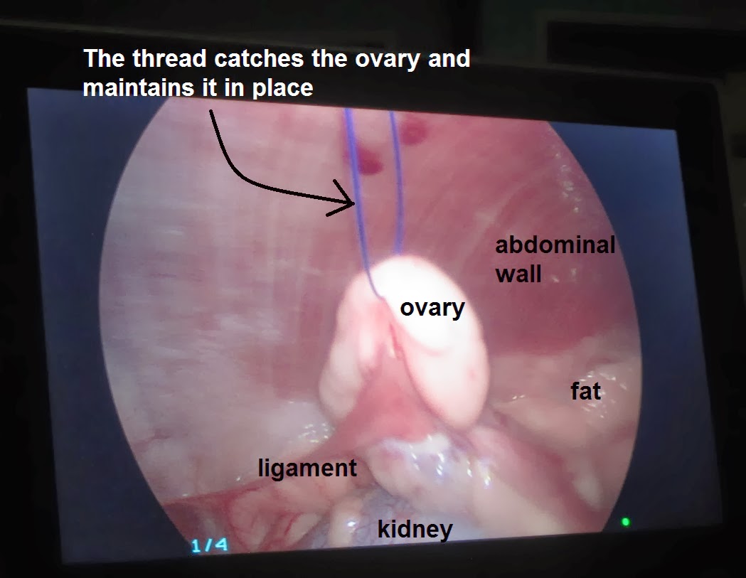
Now that we found the ovary, we would like to take it out together with its pouch (bursa) and the fat conected around it. The problem is that we only made one hole for one tool... so it would be hard to have a tool to hold the ovary properly, and another to cut it at it's base. So we use a clever technique: maintain the ovary in place by having a suture from the outside go through the wall, and hold it in place, just for the time of cutting out the ovary! :)
 |
| First grab the ovary, and put it against the wall |
 |
| Feel the location and control it on camera |
 |
| Put a stitch to grab the ovary through the wall |
 |
| See on camera that the ovary |
Now that the ovary is maintained in place, we can take out the grabbing tool (forceps) and use burner/cutter tool!
 |
| Grab, burn, cut |
 |
| Grab again, burn again, cut again |
 |
Smoke is coming out,
but it doesn't burn too much, just enough to cut safely |
 |
| One tiny tiny bit left! |
After the last snip, the ovary was free, we caught it with the forceps, undid the temporary stitch that was holding the ovary, and pulled it out through the small hole.
 |
| Grabbing the cut ovary, free the suture |
 |
| Pull it out |
 |
| There it is! |
OK, that's one side done. Then we had to do the same thing with the other ovary, using the same holes.
 |
| The tools (here the camera) is put back into position |
 |
| The table is tilted the other way, so the organs move down and away |
On the left the ovary was near the spleen, on the right, it was close to the kidney.
 |
| Burn and cut the tissue surrounding the ovary |
 |
| Pull it out and Ta Daaaaaa, the second ovary is out |
 |
| Yay we're done! |
Gastropexy
Nope! We're not done yet!
 |
| This is the stomach, try to grab it in the middle |
 |
| We cut the out part of the stomach |
 |
| And sutured it to the inner part of the abdominal wall (and first muscles) |
 |
| Compare this picture with the presurgery one. What a mess :) |
 |
| Still sleeping... |
 |
| All over! Ready to pack and go! |
Post operative care
I woke up within a few minutes. But fell back asleep. Again and again for 3 or 4 hours.
 |
| My mommy being overportective |
 |
| Amawake... whatchdishinmymoush... oh, my tongue is not working :/ |
 |
| Let's go! |
 |
| Or not...zzzzz... |
Eventually Jen and the nurse decided I am awake enough to go home safely (tram and 5 min walk).
 |
| OK I don't like this place anymore, let's go. |
I don't know exactly what happened today, but I think that was enough for one lifetime! :)